
Yolk Sac Tumor : Microcystic


Comments:
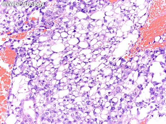
The microscopic features are similar in both pre- and postpubertal yolk sac tumors (YST). The tumor cells are arranged in a variety of architectural patterns that recapitulate the yolk sac, allantois, and extraembryonic mesenchyme. Multiple patterns coexist within the same tumor in varying proportions. Germ cell neoplasia-in-situ (GCNIS) is not present in the adjacent testis in pure YST in children. In contrast, GCNIS is a constant finding in testicular GCT in adolescents and young adults. The architectural patterns include (in order of decreasing prevalence): microcystic/reticular, myxomatous, solid, glandular/alveolar, macrocystic, endodermal sinus/perivascular, hepatoid, papillary, sarcomatoid/spindle cell, parietal, and polyvesicular vitelline pattern. Microcystic (reticular) pattern (most common): The tumor cells have large intracellular vacuoles, compressed nuclei and delicate strands of interconnected cytoplasm creating a spider-web like arrangement, frequently containing hyaline globules. In some areas, there are interconnecting ribbons and cords of tumor cells enclosing round/irregular extracellular spaces and creating a honeycomb-like meshwork. These spaces are often filled with basophilic myxoid material. Microcystic pattern often coexists and intermingles with myxomatous and solid patterns.