
About This Site
WebPathology is an educational resource with high quality pathology images of benign and malignant neoplasms and related entities. It was launched in 2003 by , with an initial focus on urologic pathology. It was subsequently expanded to include other organ systems.
The site offers high-quality specimen photographs and photomicrographs accompanied by a discussion of pertinent clinical and pathologic features. Imaging studies and management are included wherever applicable. With rapid advances in the fields of cancer genomics and precision medicine, the focus of WebPathology has recently expanded to include illustrations of molecular pathways in various cancers, including sites of targeted intervention.
The Founder
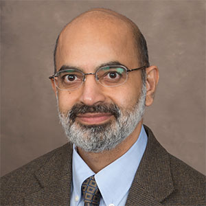 is a board-certified anatomic and clinical pathologist in private practice since 1999 in Richmond, Virginia, USA. Dr. Ramnani received his MBBS degree from Maharaja Sayajirao University in Vadodara, India, and MS (Biological Sciences) from The University of Iowa, Iowa City, IA. He completed pathology residency and surgical pathology fellowship from Univ. of Texas Southwestern Medical Center, Dallas, Texas, and urologic pathology fellowship from the Mayo Clinic, Rochester, Minnesota, before settling down in Richmond.
Dr. Ramnani has been passionate about photographing human cancers from the early days of his residency training. He has collected several hundred thousand images which he hopes to share with a world-wide audience on this site.
Acknowledgements
Numerous pathology colleagues have shared some of their interesting cases with us on WebPathology. We are grateful for their generosity. Each contribution is acknowledged in the image caption. We would especially like to thank the following pathologists for their numerous images.
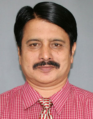
Dr. Sanjay D. Deshmukh has been a diagnostician, teacher and mentor for more than 34 years in pathology departments at several hospitals and medical colleges in Maharashtra, India. He is currently Professor of Pathology at Dr. Vithalrao Vikhe Patil Foundation’s Medical College & Hospitals, Ahmednagar, India. His areas of interest include oncopathology, hematopathology, and cytopathology. Dr. Deshmukh has published more than 70 papers.
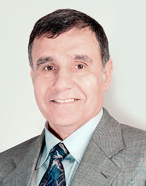 Ibrahim Zardawi, MD, is all-rounder retired senior pathologist who has worked on four continents as pathologist, educator, researcher, administrator and mentor. He was also Honorary Professor at several Australian Universities. His qualifications include, MBBS and MS (Pathology) from University of Baghdad, Iraq; FRCP (Great Britain), FRCP (Australasia), Fellow of the International Academy of Cytology, Diploma in Cytopathology (Royal College of Pathologists of Australasia), & Founding Fellow of the Faculty of Science (Royal College of Pathologists of Australasia). His peers call him “the Pathologist who loves the bit on the slide” because of his passion for pathology. He photographed most cases that came his way and has a huge collection of images. He considers Pathology as the Mother and Backbone of Medicine and that is why he wants everyone to see “Medicine through the glass slide.”
Jean-Christophe Fournet, MD, PhD, Paris, FRANCE.
Katharina Glatz-Krieger, MD, University of Basel, Basel, SWITZERLAND.
Ed Uthman, MD, Houston, Texas, USA.
Frank Gaillard, MBBS, Radiology Consultant, Royal Melbourne Hospital, Melbourne, AUSTRALIA.
Piero Picci, MD, Director, Laboratory of Experimental Oncology, Instituto Ortopedico Rizzoli, Bologna, ITALY.
Website Development Partner: Biznet, Inc., Glen Allen, Virginia, USA.
Contributing a Case to WebPathology
Please consider sharing your interesting cases with us to reach a world-wide audience. High quality gross specimen photographs are especially welcome. All contributions published on the site are appropriately acknowledged.
What to send:
- Images in JPEG or TIFF format; the larger the size the better
- A brief history – please do not include any identifying patient information
- Send them to dmr@webpathology.com
If you have any comments or suggestions about this site, please feel free to fill out the feedback form