
Parosteal Osteosarcoma

slide 72 of 93


Comments:
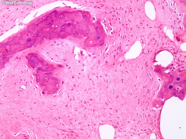
Higher magnification showing hypocellular spindle stroma separating the bony trabeculae. The stromal spindle cells lack cytologic atypia or increased mitotic activity. The vast majority of parosteal osteosarcoma are Grade I tumors; approximately 20% have sufficient atypia to be classified as Grade II.
slide 72 of 93