
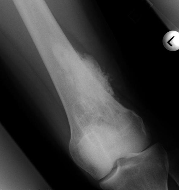
Parosteal Osteosarcoma


Comments:
Parosteal osteosarcoma is a distinct well-differentiated subtype with better prognosis than the conventional osteosarcoma. It is located on the outer surface of the cortex and is also referred to as juxtacortical osteosarcoma. The patients are slightly older (3rd and 4th decades of life) and the most commonly affected sites are metaphysis or metadiaphyseal region of femur (especially posterior distal shaft of the femur), humerus, and tibia. Radiologically, it appears densely mineralized in the center and is less radiodense at the periphery. The presence of lucency within the central portion of the lesion is usually associated with dedifferentiation. Axial CT and MRI scans are useful in confirming the surface location of the tumor and the lack of medullary involvement. Case produced with permission, courtesy of Dr. Frank Gaillard. Radiopaedia. Complete case is here.