
Steroid Cell Tumor, NOS : Microscopic

slide 52 of 74


Comments:
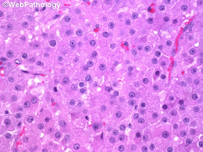
Microscopic Features of Ovarian Steroid Cell Tumor (continued from previous image): The tumor cells are medium to large-sized with distinct cytoplasmic borders. They have abundant granular eosinophilic or microvacuolated, lipid-rich cytoplasm. Yellow-brown lipochrome pigment granules are frequently seen in the cytoplasm. The nuclei are uniform and round with a prominent central nucleolus. Usually, there is no atypia and the mitotic rate is <2 mitoses/10HPF. Adjacent ovarian stroma and the contralateral ovary may show stromal hyperthecosis.
slide 52 of 74