
CMV Esophagitis

slide 73 of 74


Comments:
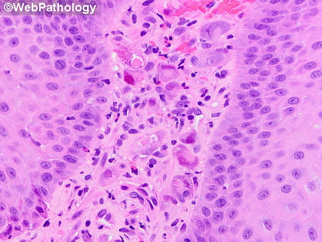
Cytomegalovirus (CMV) Esophagitis: On endoscopic esophageal biopsies, the viral inclusions are best seen in endothelial cells, stromal cells and macrophages beneath the mucosa. The infected cells are enlarged (25 to 35 microns) and show both cytoplasmic and nuclear inclusions. Cytoplasmic inclusions are basophilic or amphophilic granules that are PAS and GMS positive. Nuclear inclusions are large, round, glassy, eosinophilic structures that may be surrounded by a halo creating an owl's-eye nucleus. When the diagnosis is suspected clinically but not supported in the biopsy, immunostaining with anti-CMV monoclonal antibodies can be helpful in showing viral inclusions.Image courtesy of: Phoenix Bell, MD.
slide 73 of 74