
Herpes Esophagitis

slide 20 of 74


Comments:
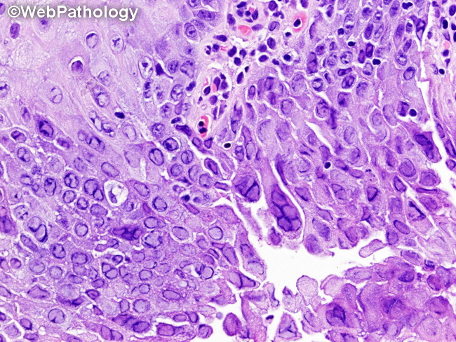
Herpes Esophagitis: Higher magnification of the previous image from the edge of an ulcer shows numerous squamous epithelial cells (including a few multinucleated cells) with ground glass nuclei and marginated chromatin. The intranuclear inclusions may not always be identifiable, especially if the biopsy is taken from the center of the ulcer instead of the margins.
slide 20 of 74