
Herpes Esophagitis

slide 19 of 74


Comments:
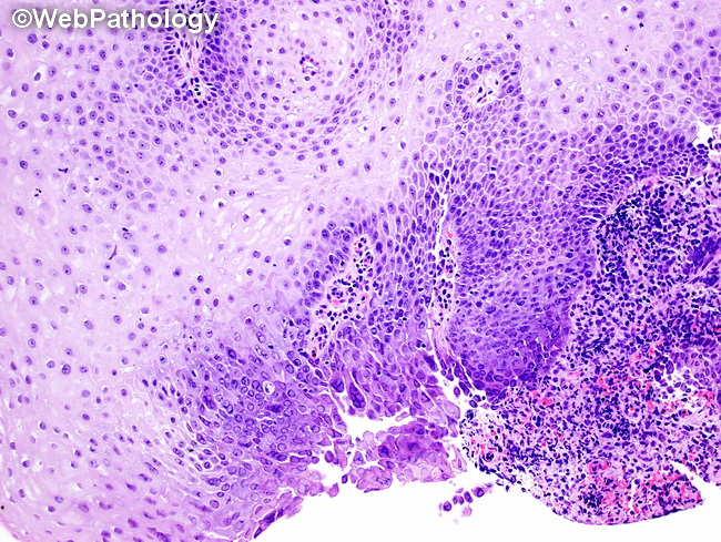
Herpes Esophagitis: The characteristic histologic findings in endoscopic esophageal biopsies include: multinucleated cells with nuclear molding and chromatin margination; large, eosinophilic, ground glass intranuclear inclusions with a halo (Cowdry A bodies); and a background of acute inflammation with ulceration. The viral cytopathic effects are best seen in viable squamous epithelial cells at the margins of the ulcer. They contain intact and fragmented virions. The presence of HSV can also be confirmed by PCR, in-situ hybridization and immunohistochemistry.
slide 19 of 74