
Mixed Epithelial & Stromal Tumor

slide 19 of 28


Comments:
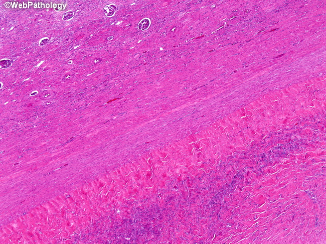
The lower right portion of the image shows periphery of the tumor composed of dense fibroblastic stroma resembling ovarian stroma that is sharply demarcated from the normal kidney (upper left). The patient was a 69 y/o female who presented with abdominal mass. She was found to have a multicystic lesion in her left kidney and underwent nephrectomy. The final diagnosis was mixed epithelial and stromal tumor of the kidney.
slide 19 of 28