
Ovary : Cellular Fibroma


Comments:
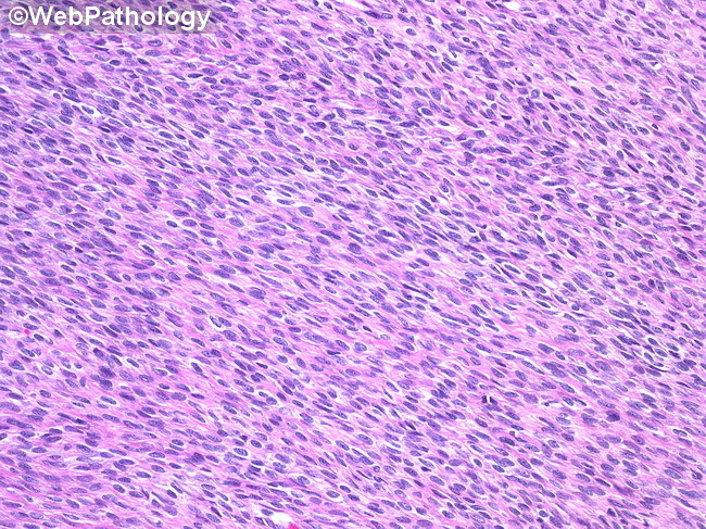
Microscopic Features: Cellular fibromas are composed of densely cellular areas admixed with less cellular areas of conventional fibroma. Stromal edema and hyalinization are less common in these tumors. Areas of infarct-type geographic necrosis that are sharply demarcated from viable areas may be present. In such areas, concentric zones of viable tumor are present around blood vessels. The cells are oval or spindle-shaped and are arranged in short intersecting bundles or in a storiform pattern. They have round to oval nuclei with minimal atypia and 1-3 mitoses/10HPF. Cellular fibromas with higher mitotic rates (4-20 mitoses/10HPF) but without cytologic atypia are called mitotically-active cellular fibromas. Both cellular and mitotically-active cellular fibromas frequently show trisomy 12.