
Hibernoma : References


Comments:
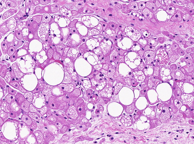
References:1. Goldblum, J. R., Weiss, S. W., & Folpe, A. L. (2020). Enzinger & Weiss's Soft Tissue Tumors - 7th Edition. Philadelphia, PA. Elsevier.2. Soft Tissue and Bone Tumours. WHO Classification of Tumours (WHO Fascicle). 5th Edition 2020.3. Goldblum, J. R. et al (2018). Rosai and Ackerman's Surgical Pathology - 11th Edition. Philadelphia, PA. Elsevier.4. Calonje, E et al (2020). McKee's Pathology of the Skin with Clinical Correlations - 5th Ed. Elsevier.This image of a hibernoma shows an admixture of: 1) large polygonal, round or ovoid cells with abundant granular eosinophilic cytoplasm, distinct cytoplasmic borders, and round centrally-placed nuclei; 2) multivacuolated large adipocytes with central nuclei; and 3) mature univacuolated adipocytes (white fat). There is no cytologic atypia and mitotic activity is not increased.