
Diagnosis of Gout : Urate Crystals in Synovial Fluid


Comments:
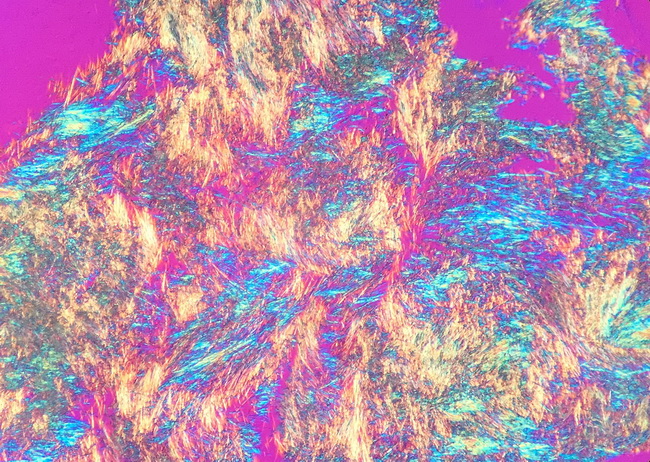
Monosodium urate (MSU) in Joint Fluid & Tophi: The diagnosis of gout rests on demonstration of MSU crystals in joint fluid or in tophi. Patients with new-onset acute monoarticular arthritis must undergo arthrocentesis with examination of synovial fluid for crystals. It is important to rule out septic arthritis in these patients because, if left untreated, it can rapidly destroy the joint. A prior history of gout or pseudogout does not exclude septic arthritis. On the contrary, septic arthritis commonly develops as a complication in crystal-induced arthritis. Tophi can also be aspirated for crystal analysis under polarizing microscopy. The joint fluid should be sent for cell count and differential, gram stain, culture and sensitivity, and microscopic analysis for crystals. MSU crystals are needle-shaped and exhibit negative birefringence in polarized light, i.e. when examined with a polarizing filter and red compensator filter, they are yellow when aligned parallel to the slow axis of the red compensator but appear blue when aligned across the direction of polarization. They are seen in both intracellular and extracellular locations. Serum Urate Levels: Serum uric acid levels are unreliable for the diagnosis of gout. They may be normal during an acute attack and hyperuricemia does not prove the diagnosis of acute gout. Image courtesy of: Ed Uthman, MD, Houston, TX.