
Osteoblastoma

slide 3 of 14


Comments:
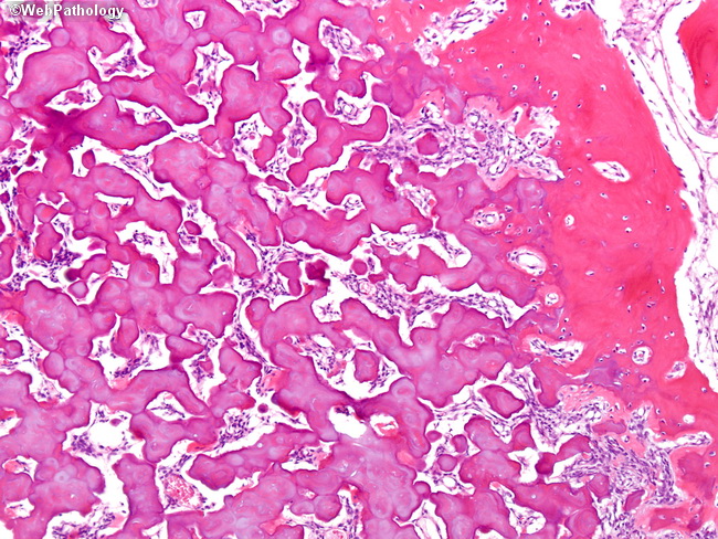
The image shows anastomosing bony trabeculae separated by loose fibrovascular stroma. Towards the periphery of osteoblastomas, the neoplastic bony trabeculae merge with those of the host bone giving rise to an appearance of maturation (right side of the image). Osteoblastomas show remarkable male predominance. Most cases occur within the first three decades of life. The bones most often involved include vertebral column (especially the posterior elements) and the long bones.
slide 3 of 14