
Osteoblastoma : Radiographic Features

slide 11 of 14


Comments:
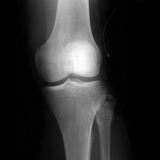
On plain films, osteoblastomas usually appear as expansile lytic lesions with a sclerotic rim. The location of tumor in this case is the upper end of fibula. There is a hint of internal calcification. Some cases show an associated soft tissue mass. Secondary aneurysmal bone cyst changes are seen in about 20% of cases. Case produced with permission, courtesy of Dr. Frank Gaillard. Radiopaedia. Complete case is here.
slide 11 of 14