
Gallstones : Radiology

slide 9 of 73


Comments:
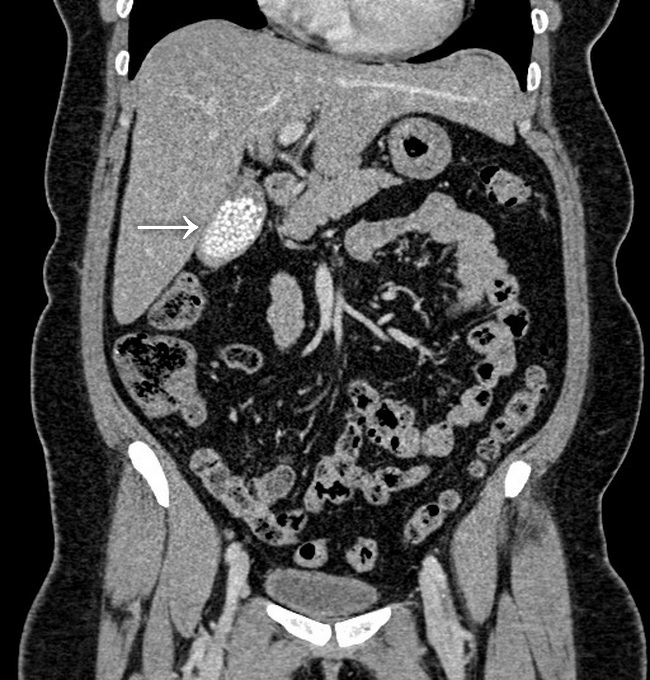
This abdominal CT with contrast (coronal view) is from a 50 y/o female who presented with abdominal pain. The CT shows numerous small gallstones within a thin-walled gallbladder (money-bag appearance). Case courtesy of Ian Bickle, Radiopaedia.org. From the case rID: 60354; used under Creative Commons License
slide 9 of 73