
Brown Pigment Gallstones : Pathophysiology


Comments:
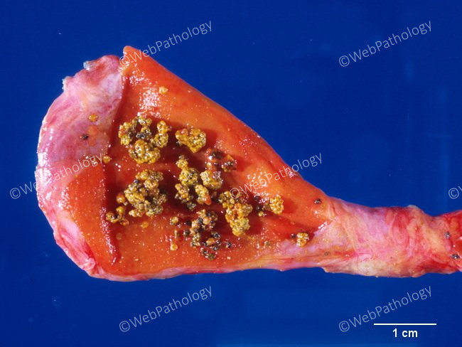
Pathophysiology of Brown Pigment Gallstones: Brown gallstones usually form in infected or obstructed bile ducts and sometimes within the gallbladder. Organisms reach the biliary tract via blood stream from a focus of infection (tonsils, bowel, dental tartar and caries) or from the bowel via lymphatics. Biliary stasis caused by infection plays a major role. Organisms implicated include bacteria (E. coli, actinomyces, typhoid bacilli) and parasites (Clonorchis sinensis, Opisthorchis viverrini, and Ascaris lumbricoides). The exfoliated mucosal cells, bacterial debris, parasitic ova, and even foreign bodies like ingested vegetable debris (in rare instances) provide the nidus around which the stones is formed. Moynihan's Aphorism (attributed to the English surgeon Lord Moynihan of Leeds (1865-1936): 'A gallstone is a tombstone erected to the memory of the organism within it'. (pathophysiology continues in the next image)