
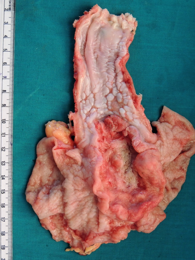
Adenocarcinoma of Gastro-Esophageal Junction


Comments:
High-grade adenocarcinoma arising at the gastro-esophageal (G-E) junction in a 68 y/o female. The patient presented with one-month history of progressive dysphagia to solid foods and regurgitation. Endoscopic and contrast radiographic studies revealed an ulcerated lesion at the G-E junction. Following an endoscopic biopsy confirmation of the diagnosis, the tumor was resected. The resected specimen consists of distal 11 cm segment of the esophagus and an 8 cm portion of cardiac end of the stomach. The G-E junction shows a 5 cm x 4 cm ulcerated mass with everted margins and necrotic material in its floor. Some of the celiac lymph nodes were positive for metastatic tumor. Case courtesy of: Dr. Sanjay D. Deshmukh, Professor of Pathology, Dr. Vithalrao Vikhe Patil Medical College & Hospitals, Ahmednagar, India.