
Hairy Leukoplakia


Comments:
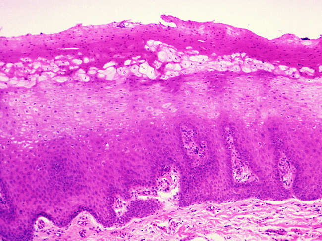
Hairy Leukoplakia: Microscopically, there is acanthosis and parakeratosis with surface corrugations. Beneath the surface, in the upper spinous layer, there is a band of vacuolated and balloon cells with abundant pale cytoplasm. The pale cells show nuclear clearing (Cowdry type A inclusions) with peripheral condensation of chromatin in a beaded pattern against the nuclear membrane. There is no dysplasia. Candida hyphae are found in almost 80% of cases, albeit without the inflammatory response. In the right clinical context, the diagnosis is easily made on macroscopic and microscopic appearance. Special techniques, including immunohistochemistry, PCR, and in-situ hybridization are available to confirm the presence of EBV within keratinocyte nuclei if necessary. Image credit: Sol Silverman, Jr., DDS; CDC/PHIL.