
Choledochal Cyst : Microscopic Features


Comments:
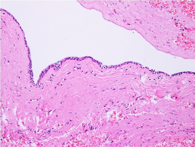
Microscopic Features of Choledochal Cyst: The appearance depends upon the patient's age. Cysts excised from infants have intact columnar epithelium and scant inflammation. With advancing age, the epithelial lining is often partially or completely destroyed by inflammation and fibrosis. Foci of dystrophic calcification may be present. Smooth muscle is often present in the wider portions of the cyst. There may be associated chronic cholecystitis. Intestinal metaplasia is frequently seen in residual epithelium in choledochal cysts removed from patients older than 15 years. Goblet cells, Paneth cells, and neuroendocrine differentiation has also been reported.This choledochal cyst was removed from a 57 y/o female who presented with abdominal pain. The cyst is partially lined by biliary type epithelium. The fragment near the top is devoid of epithelial lining. No smooth muscle was present. Image source: Hwaseong Ryu et al. A Rare Case of Extrahepatic Left Hepatic Duct Diverticulum: Case Report with Literature Review. Korean J Pancreas Biliary Tract 2019; 24(1): 31-34; DOI: https://doi.org/10.15279/kpba.2019.24.1.31; image used under Creative Commons Noncommercial Attribution License.