
Intraductal Papillary Neoplasm of Bile Duct : Macroscopic


Comments:
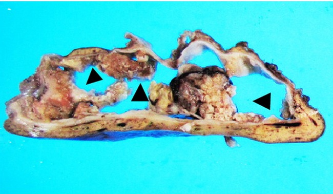
Macroscopic Appearance of Intraductal Papillary Neoplasm of Bile Ducts (IPNB): (continued from previous two images). Multilocular cystic forms of IPNB do occur (15% cases). The cysts are filled with mucinous secretions covering intracystic papillary lesions. The inner lining is smooth or granular. IPNBs arising in peribiliary glands also appear as intracystic papillary lesions around the large bile ducts. The areas of stromal invasion may be grossly inconspicuous or form solid, ill-defined areas around the papillary tumor. There are geographic differences in macroscopic appearance. In Asian patients, IPNBs more often involve intrahepatic bile ducts and secrete copious mucin. In Western countries, extrahepatic bile ducts or hepatic hilum are more commonly involved and mucin hypersecretion is rare.The image shows cystic IPNB. There are multiple polypoid mural nodules lining a thin-walled cyst which communicated with the bile duct. Image source: Ohtsuka M. et al. Intraductal Papillary Neoplasms of the Bile Duct. International Journal of Hepatology Volume 2014, Article ID 459091, 10 pages http://dx.doi.org/10.1155/2014/459091; image cropped from the original and used under Creative Commons Attribution License.