
Lipoma Arborescens : Diagnosis


Comments:
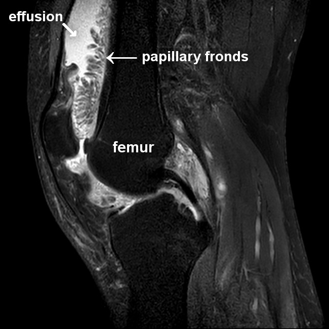
Diagnosis of Lipoma Arborescens: Imaging studies (CT, MRI, ultrasound) can effectively make the diagnosis. MRI is the diagnostic modality of choice. On MRI, it shows multiple, frond-like villi projecting into the joint space from the synovium. It has high signal intensity on T1- and T2-weighted images similar to subcutaneous fat and a low signal on fat sat series (as shown in this sagittal view). Not infrequently, there are other associated joint lesions, including joint effusion, degenerative changes, meniscal tears, synovial cysts, synovial chondromatosis, and bone erosions. Joint fluid aspiration may become necessary to look for urate crystals and microorganisms to rule out other causes of joint swelling. Case courtesy of Bassem Marghany, Radiopaedia.org. From the case rID: 75123