
Amebic Colitis

slide 19 of 23


Comments:
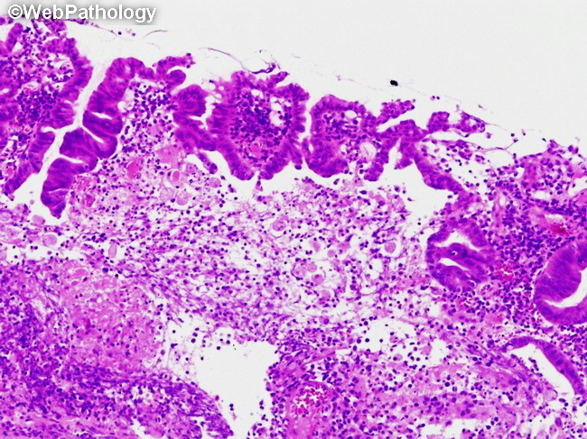
Histologic examination of a colonic biopsy taken at the edge of an ulcer shows variable non-specific inflammatory infiltrate. There are flask-shaped ulcers with a narrow neck and broad base. The presence of amebic trophozoites confirms the diagnosis. This colonic biopsy image is from an immunocompromised patient with amebic dysentery. Image courtesy of: @Patholwalker
slide 19 of 23