
Amebic Colitis

slide 18 of 23


Comments:
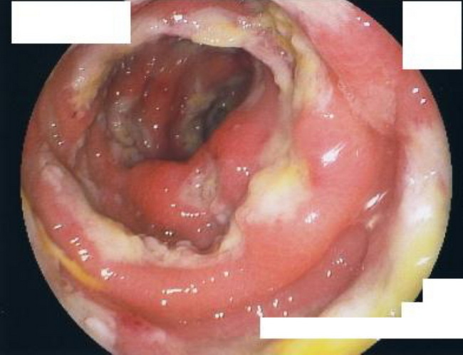
The gold standard for the diagnosis of amebic colitis is colonoscopy with biopsy. Since cecum and ascending colon are most often involved, colonoscopy is preferred over sigmoidoscopy. The classic appearance consists of multiple punctate ulcers measuring 2 to 10 mm and covered with exudate. In severe cases, the ulcers may coalesce producing large necrotic areas with normal intervening mucosa. This endoscopic view is from an immunocompromised patient with amebic dysentery who eventually underwent colectomy. Image courtesy of: @Patholwalker
slide 18 of 23