
MSI-High Colorectal CA : Morphology


Comments:
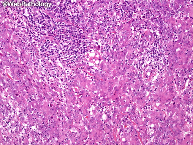
MSI-high colorectal carcinomas are more likely to be poorly-differentiated or show mucinous differentiation. The growth pattern in poorly-differentiated tumors consists of solid sheets, trabeculae, nests, and organoid areas with pushing expansive borders instead of infiltrating borders. Gland formation is absent or rare. The tumor cells are polygonal with moderate amount of eosinophilic or amphophilic cytoplasm, uniform vesicular nuclei and prominent nucleoli. Mitotic activity is increased. Geographic necrosis or comedonecrosis may be present. There is no �dirty necrosis� seen in conventional colorectal adenocarcinoma. Small areas (<20%) of well-differentiated conventional adenocarcinoma or mucinous carcinoma may be present. One of the prominent features is the presence of numerous tumor-infiltrating lymphocytes. The edge of the tumor shows lymphoid aggregates or follicles with or without germinal centers.