
FOXO1 Breakapart FISH Probe

slide 60 of 94


Comments:
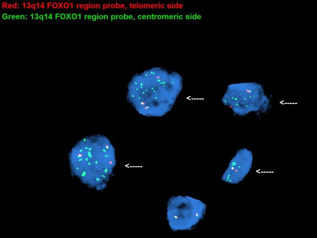
This image of FOXO1 breakapart FISH probe from a case of alveolar rhabdomyosarcoma shows PAX7-FOXO1 gene rearrangement associated with t(1;13)(p36;q14) translocation (arrows). In addition, there is amplification of the fusion gene - a feature more likely seen with PAX7-FOXO1 fusion than PAX3-FOXO1 fusion. The cell at the bottom of the image is a normal interphase cell without translocation at 13q14.11 band involving the FOXO1 gene. It shows two green/red signals that co-localize as pairs and represent two non-rearranged 13q14.11 loci. Image courtesy of: Henry Tran, MD; used with permission.
slide 60 of 94