
Dysplasia in Ulcerative Colitis

slide 38 of 60


Comments:
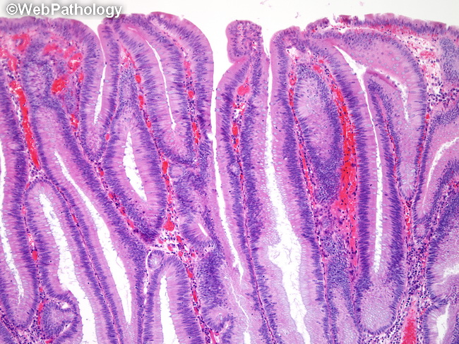
This image shows a tubulo-villous adenoma with low-grade dysplasia discovered during surveillance colonoscopy in a 60-year old male with 10+ year history of ulcerative colitis. The tubulo-papillary fronds are lined by uniform cells that maintain their polarity. The nuclei are slightly enlarged, elongated, and hyperchromatic. There is nuclear overcrowding and pseudostratification. Nucleoli are not prominent. Mature goblet cells are reduced in number. Architecturally, the glands show crowding and slight irregularity.
slide 38 of 60