
Synovial Sarcoma : Differential Diagnosis


Comments:
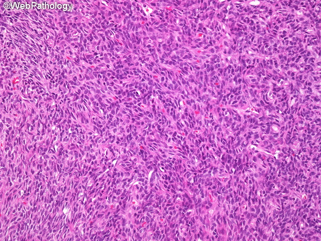
Differential Diagnosis of Monophasic Synovial Sarcoma: Monophasic fibrous synovial sarcoma (shown here) has to be distinguished from other spindle cell neoplasms, including fibrosarcoma, leiomyosarcoma, MPNST, solitary fibrous tumor/hemangiopericytoma, and spindle cell carcinoma. Fibrosarcoma: Features favoring synovial sarcoma include location near a major joint, multilobular growth pattern, plump nuclei, whorled architectural pattern of spindle cells, mast cells, calcification, and focal hemangiopericytomatous vasculature (stag-horn vessels). Other findings supporting synovial sarcoma include positivity for EMA, cytokeratin, and TLE1 in the tumor cells and the presence of chromosomal translocation t(X;18)(p11.2;q11.2) or SS18/SSX hybrid protein. Leiomyosarcoma: Features favoring leiomyosarcoma over monophasic fibrous synovial sarcoma include: arrangement of spindle cells in fascicles intersecting often at right angles, blunt-ended nuclei with paranuclear vacuole, densely eosinophilic cytoplasm, and strong immunoreactivity for one or more muscle markers (smooth muscle actin, muscle-specific actin, desmin, and h-caldesmon). MPNST: MPNST may be seen clearly originating from a nerve or arise from a preexisting neurofibroma or be seen in patients with neurofibromatosis 1. The spindle tumor cells of MPNST appear wavy or buckled. Immunohistochemically, MPNSTs are positive for S-100 (at least focally, in two-thirds of cases) and HMGA2 and negative for CK7 and CK19. Synovial sarcomas are S-100 positive in one-third of cases, positive for CK7 and CK19, and negative for HMGA2. Rare examples of synovial sarcoma arising from peripheral nerves have been reported. Solitary Fibrous Tumor/Hemangiopericytoma: Focal hemangiopericytomatous vascular pattern (stag-horn vessels) may be seen in synovial sarcoma. Distinction from solitary fibrous tumor/hemangiopericytoma can be made with immunohistochemical and molecular genetic studies. Synovial sarcomas express epithelial markers and TLE1 and are negative for CD34. SFT has opposite staining pattern.