
Pineocytoma


Comments:
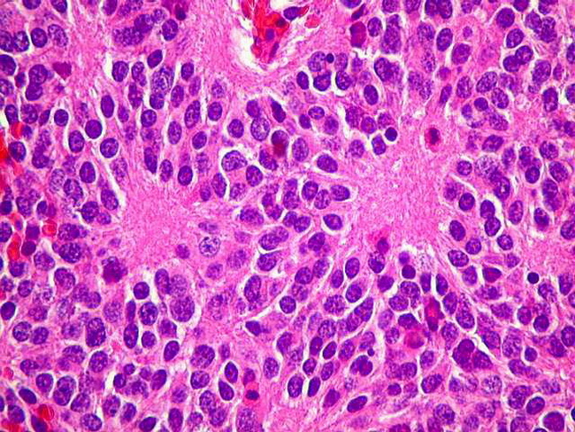
Pineocytomas are WHO Grade I tumors that make up between 15% and 30% of pineal parenchymal tumors. The peak incidence is seen between 25 to 35 years of age. The presenting symptoms are related to increased intracranial pressure, visual disturbances, or hypothalamic dysfunction. Grossly, they are well-demarcated lesions which may have areas of cystic degeneration or focal necrosis. They are composed of small uniform cells with round nuclei resembling mature pineocytes. They contain pineocytomatous rosettes (which resemble Homer-Wright rosettes) with abundant neuropil at the center surrounded by uniform neoplastic cells. Courtesy of: Dr. Luciano de Souza Queiroz, Dept. of Pathology, Faculty of Medical Sciences, State University of Campinas (UNICAMP), Campinas, S�o Paulo State, BRAZIL. Additional images are here.