
Papillary Craniopharyngioma

slide 29 of 32


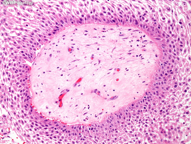
Comments:
Higher magnification showing well-differentiated non-keratinizing squamous epithelium arranged around a fibrotic stromal core. Note the dehiscence of epithelium at the peripheral portions which results in pseudopapillae formation. The mature squamous cells retain their nuclei (unlike the anucleated squames of adamantinomatous craniopharyngioma). Wet keratin is not seen. Peripheral palisading is not as prominent as in adamantinomatous subtype.
slide 29 of 32