
Keratoacanthoma : Clinical Presentation


Comments:
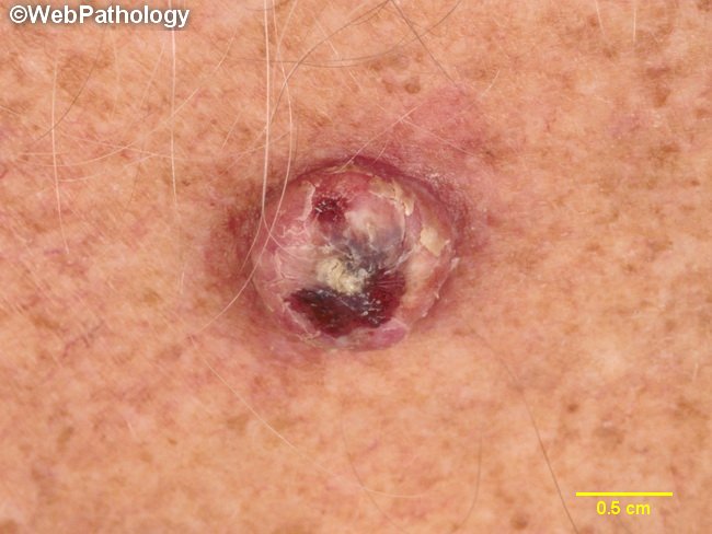
Keratoacanthoma is a tumor of the infundibular portion of the hair follicle but closely resembles conventional squamous cell carcinoma of the skin and is therefore presented in this section. Some consider it to be a distinct variant of squamous cell carcinoma. Clinical Variants:Solitary Keratoacanthoma: It is more common in light-skinned races and is due to excessive exposure to UVB rays. It occurs predominantly on sun-damaged areas of face, forearms, wrists, and back of hands. Rare locations include lips, oral mucosa, tongue, vulva, perianal skin, and penis. Some cases are associated with inflammatory dermatoses, congenital skin lesions, genetic disorders, or scars of previous trauma. Most patients are in 6th or 7th decade and it is more common in males (M:F = 4:1). The lesion appears as a smooth, dome-shaped papule that rapidly grows over 4-6 weeks into a 1-2 cm diameter round flesh-colored nodule with a central crater filled with keratinaceous debris. Rare cases may be several cm large (giant keratoacanthoma) or have multiple lesions. A typical case usually shows spontaneous regression after a period of initial growth leaving behind a depressed hypopigmented scar. The total duration of the lesion being 4-6 months. Clinical variants - continued in the next slide.