
Chronic Tophaceous Gout : Gross Pathology


Comments:
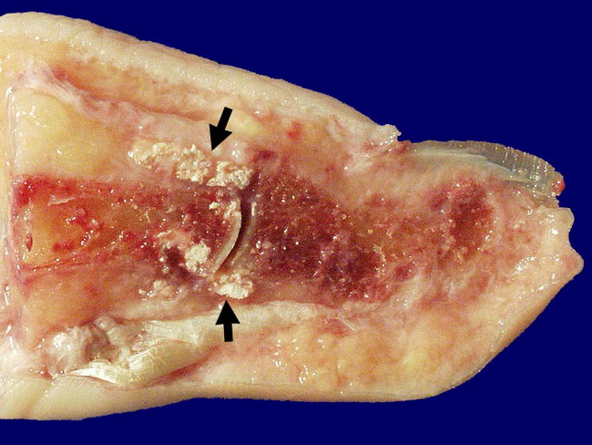
Multiple cycles of monosodium urate (MSU) crystal deposition during acute attacks eventually lead to chronic tophaceous arthritis. Formation of tophi is hallmark of chronic gout. Chalky yellow-white deposits of MSU form on the articular surface, synovium, ligaments, tendons, bursae, and periarticular soft tissues. Uncommonly, tophi may be seen in earlobes and fingertips. Superficial urate deposits may even ulcerate through the skin discharging chalky-white material. The synovium becomes thickened and shows chronic inflammation and fibrosis. There is erosion of articular cartilage and irregular destruction of juxta-articular bone. Fibrosis and ankylosis severely limit the joint mobility and function. Advanced cases with large gouty tophi are rarely seen nowadays due to advances in the diagnosis and treatment of the disease. The image shows white gout crystal deposits in the soft tissues and bones of the proximal interphalangeal joint of the big toe. Image copyright: pathorama.ch