
Synovial Sarcoma : Radiology


Comments:
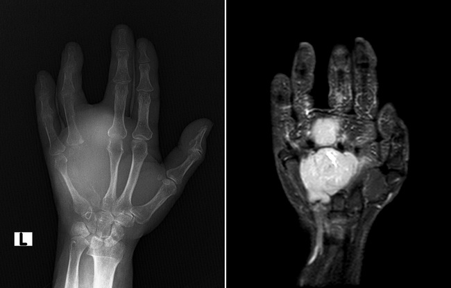
Almost 85% to 95% of synovial sarcomas arise in the extremities, especially around the knee joint followed by foot, lower leg-ankle region, and hip-groin area. Tumors arising in upper extremities are evenly distributed among wrist, elbow, shoulder, and hand. Case History: The patient was an elderly female who presented with swelling of hand of a few months duration. Plain radiograph (left panel) shows near total erosion of the fourth metacarpal bone with soft tissue tumefaction. There is widening of space between the third and fifth metacarpal bones extending distally in between the middle and ring fingers. MRI with contrast (right panel) shows a 7.1 x 5.1 x 4.6 cm lobulated ill-defined soft tissue mass in the left hand. The mass is seen eroding the fourth metacarpal bone for a 3.6 cm segment and widening the space between the third and fourth fingers. The final diagnosis was synovial sarcoma. Case courtesy of Dr Henry Knipe, Radiopaedia.org. From the case rID: 63840