
Osteosarcoma : Radiographic Features

slide 11 of 93


Comments:
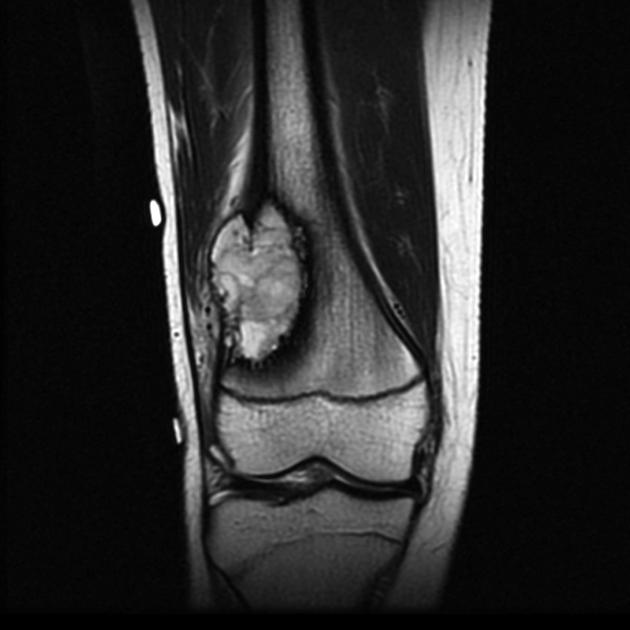
Magnetic resonance imaging (MRI) has become an essential tool in osteosarcoma for accurate local staging, evaluation of intramedullary extension of the tumor, soft tissue involvement, and assessment of the growth plate. This information is vital in planning for limb-sparing surgery. In T2 weighted images, the non-mineralized soft tissue components have high signal intensity as depicted here (same case as the previous two images). Case produced with permission, courtesy of Dr. Frank Gaillard. Radiopaedia. Complete case is here.
slide 11 of 93