
Barrett Esophagus : Introduction


Comments:
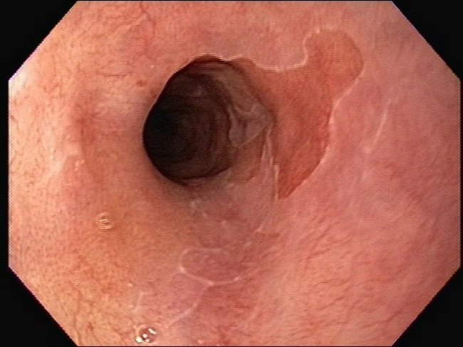
Introduction: Barrett esophagus (BE) is a metaplastic change characterized by replacement of the normal stratified squamous epithelium of the distal esophagus with columnar epithelium containing goblet cells (intestinal metaplasia). It is named after Norman Barrett, an Australian surgeon who described it in 1950's. BE arises secondary to chronic gastroesophageal reflux disease (GERD), in which acid refluxes from the stomach into the lower esophagus. Over time, the acid-damaged squamous epithelium is replaced by columnar epithelium which is better equipped to handle an acidic environment through its mucin secretions and tight junctions. The columnar epithelium by itself causes no symptoms. The presence of BE is a risk factor for the development of dysplasia and ultimately, esophageal adenocarcinoma (EAC). The pathogenesis of BE remains unknown. This endoscopic image shows a small patch of orange-pink (salmon-colored) mucosa in the distal esophagus. When < 3cm of metaplalstic epithelium lines the distal esophagus (as in this case), it is categorized as short-segment BE. Image courtesy of: Pramod Malik, MD; used with permission.