
Warthin's Tumor

slide 77 of 110


Comments:
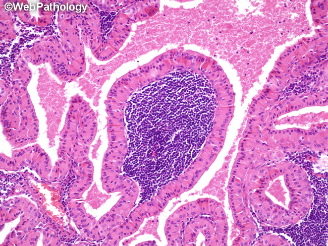
High power view of Warthin�s tumor showing papillary structures lined by bi-layered oncocytic epithelium composed of tall columnar luminal cell layer and a discontinuous layer of basally locatedcuboidal cells. The stroma shows dense lymphocytic infiltrate with germinal center formation. The cystic spaces are filled with eosinophilic granular material.
slide 77 of 110