
Pindborg Tumor : Radiology


Comments:
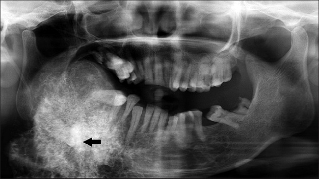
On plain films, calcifying epithelial odontogenic tumor appears as a unilocular (more frequent in maxilla) or multilocular, radiolucent or mixed radiolucent/radio-opaque lesion with often well-defined scalloped margins. About 20% of cases have ill-defined margins. The tumor is frequently associated with an impacted molar (usually mandibular molar). The appearance of calcified streaks emanating from a central radio-opaque lesion has been likened to a "driven-snow" pattern. This panoramic radiograph is from the same case as the previous three images. It shows a single well-defined circular mixed radiopaque-radiolucent lesion on the right side of the mandible with displacement of the mandibular and maxillary teeth. Presence of a radiopaque mass in the center of the lesion (black arrow) with radiopaque streaks has the appearance of "driven snow". Image source: Misra et al. Giant Pindborg Tumor (Calcifying Epithelial Odontogenic Tumor) : An Unusual Case Report with Radiologic-Pathologic Correlation. J Clin Imaging Sci 2013, 3:11. Used under the terms of Creative Commons Attribution-NonCommercial-ShareAlike 4.0 License.