
Cutaneous Mastocytoma : Gross
Home
 Hematopathology
Myeloid, Histiocytic & Dendritic Cell Neoplasms
Mastocytosis
Cutaneous Mastocytoma : Gross
Hematopathology
Myeloid, Histiocytic & Dendritic Cell Neoplasms
Mastocytosis
Cutaneous Mastocytoma : Gross

slide 30 of 64


Comments:
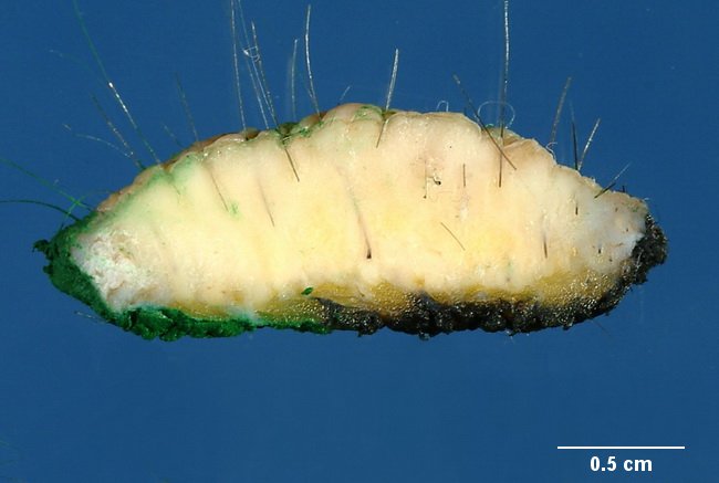
Cut surface of a resected specimen of cutaneous mastocytoma (same case as the previous image). The normal architecture is replaced by a diffuse, yellow-orange infiltrate filling up dermis and extending into subcutaneous tissue, corresponding to sheets of mast cells seen microscopically. Image courtesy of Dr. Jean-Christophe Fournet, Paris, France; humpath.com; used with permission.
slide 30 of 64