
Urticaria Pigmentosa : Microscopic


Comments:
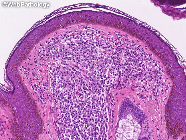
Microscopic Features of Urticaria Pigmentosa (continued from previous slide): The neoplastic mast cells are histologically similar to normal mast cells. They are oval, polygonal, or spindle shaped and have moderately abundant eosinophilic or amphophilic cytoplasm with faintly visible granules. The nuclei are round to oval with clumped chromatin and inconspicuous nucleoli. The nuclei are centrally located and create a "fried egg" appearance. Binucleated or multinucleated cells may be present. Cytologic atypia is not a common feature. Adult lesions tend to have fewer mast cells than those in children and may overlap with reactive changes that may be seen in normal or inflamed skin. If the skin lesions have been scratched or picked upon before biopsy, the papillary dermis may show edema as well as lymphocytes, histiocytes, and eosinophils. Older lesions may show basal cell hyperpigmentation in the overlying epidermis.