
Urticaria Pigmentosa : Microscopic
Home
 Hematopathology
Myeloid, Histiocytic & Dendritic Cell Neoplasms
Mastocytosis
Urticaria Pigmentosa : Microscopic
Hematopathology
Myeloid, Histiocytic & Dendritic Cell Neoplasms
Mastocytosis
Urticaria Pigmentosa : Microscopic

slide 14 of 64


Comments:
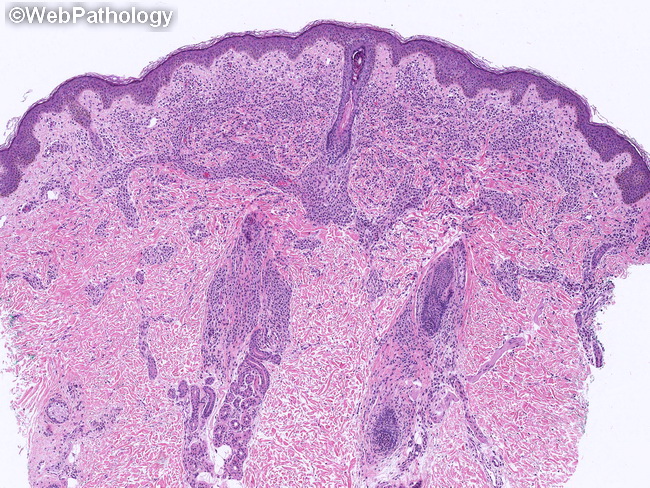
Microscopic Features of Urticaria Pigmentosa: The skin biopsies show sheets or aggregates of uniform-appearing neoplastic mast cells which are histologically indistinguishable from normal mast cells. The diagnosis of mastocytosis depends upon their number, distribution, and immunohistochemical profile. The mast cells fill the papillary dermis and extend into reticular dermis in a perivascular and periadnexal distribution. microscopic features continued in the next slide.
slide 14 of 64