
LCH of Bone : Differential


Comments:
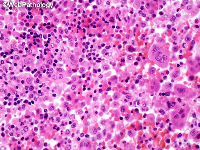
Differential Diagnosis of Langerhans Cell Histiocytosis of Bone: Early bone lesions can raise the concern for malignancy due to higher cellularity, infiltrative nature, bone destruction, and soft tissue extension. The differential includes Ewing sarcoma, osteosarcoma, and Hodgkin lymphoma. Bone lesions with pathologic fractures, hemorrhage, or necrosis can show xanthogranulomatous inflammation and may be mistaken for fibrohistiocytic lesions. Late (healing) lesions show extensive sclerosis, lymphoplasmacytic infiltrate, and sparse or absent Langerhans cells. They may resemble chronic osteomyelitis. The clinical picture in LCH of mandible or maxilla can mimic severe periodontitis. The image shows a few osteoclast-type giant cells admixed with Langerhans cell histiocytes, eosinophils, neutrophils, lymphocytes, and hemorrhage.