
LCH of Bone : Morphology


Comments:
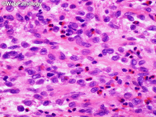
Langerhans Cell Histiocytosis (LCH) of Bone: The lytic bone lesions of LCH show a diffuse infiltration of Langerhans cells which are large, pale-staining mononuclear cells resembling histiocytes. They have indistinct cytoplasmic borders and complex, grooved or folded nuclei. The background consists of variable number of eosinophils, osteoclast-type giant cells, macrophages, and a few T-lymphocytes. Plasma cells are usually sparse or absent. In some cases, hemorrhage, necrosis, and neutrophilic infiltrate can be prominent. Immunohistochemical Profile: As in other sites, Langerhans cells are positive for langerin (CD207) in a granular cytoplasmic pattern and CD1a (membranous and paranuclear staining). S100 protein positivity is not as useful in bone lesions because it stains chondrocytes as well.