
Pulmonary LCH : Clinical


Comments:
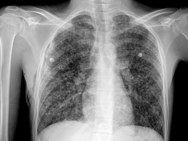
Pulmonary Langerhans Cell Histiocytosis (LCH) : It is a special type of LCH and was previously referred to as eosinophilic granuloma, histiocytosis X, or Langerhans cell granulomatosis. It is an interstitial lung disease that is usually seen in adults in 3rd or 4th decades of life. It occurs almost exclusively in smokers (>90% of cases) and may spontaneously regress upon cessation of smoking. About 20% of patients have extrapulmonary manifestations such as bone or pituitary lesions. Less commonly, pulmonary LCH occurs in children as a component of multisystem disseminated disease. Two-thirds of the patients are asymptomatic and the disease is discovered incidentally on imaging studies performed for other reasons; the remainder present with non-productive cough, dyspnea, or spontaneous pneumothorax. Most patients go into remission upon cessation of smoking. Some develop progressive disease that often proves fatal. Pulmonary LCH shows diffuse involvement of lungs with predilection for upper lobes as seen in this plain chest x-ray (frontal view) from a 20 y/o male who presented with acute breathlessness. There are bilateral diffuse cystic changes with associated reticulonodular shadows. Case courtesy of Dr Kashif Nadeem Liaqat, Radiopaedia.org. From the case rID: 29900