
Sezary Syndrome : Microscopic Features


Comments:
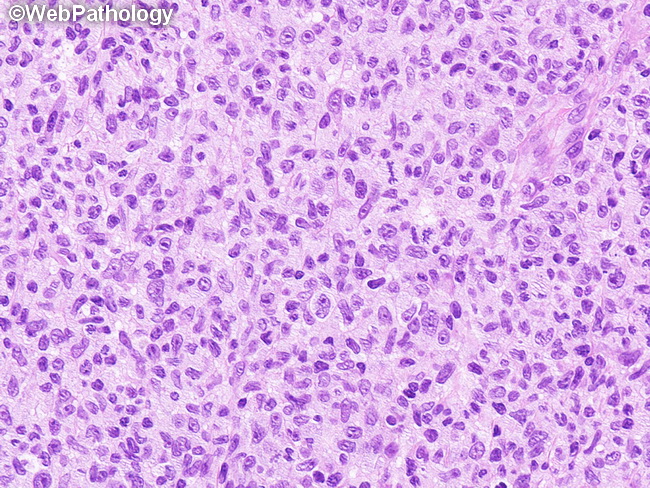
Sezary syndrome (SS) - Microscopic Features (continued from previous slide): In as many as 30% to 40% of patients with classic clinical picture of SS, the histologic findings in skin biopsies may non-diagnostic or non-specific and only show spongiosis. The reasons for non-diagnostic biopsies include inadequate sampling as well as use of topical therapy prior to biopsy. If Sezary syndrome is in the differential diagnosis, multi-color flow cytometry on the peripheral blood is a more reliable tool. Lymph nodes involved in SS show features of dermatopathic lymphadenopathy. There may be partial or complete effacement of the nodal architecture by Sezary cells. This high magnification image of a skin biopsy shows small, medium-sized, and a few large lymphoid cells with convoluted nuclei.