
Sezary Syndrome : Immunophenotype


Comments:
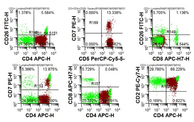
Sezary syndrome (SS) - Immunophenotype: Multicolor flow cytometry on peripheral blood, in conjunction with detection of clonal TCR gene rearrangement by PCR, is a reliable tool for the diagnosis of Sezary syndrome. The neoplastic cells are mature helper T-cells that are usually CD2+, CD3+, CD4+ CD5+, CD7+ and CD8-ve. As in mycosis fungoides, aberrant loss of pan-T-cell antigens, including CD2, CD3, CD4, CD5, CD7, and CD26 is frequently seen. Of these, aberrant loss of CD7 (≥ 40% of cells) and/or CD26 (≥ 80% of cells) is highly sensitive and specific for the diagnosis of SS. Multi-color flow cytometry could also be used to demonstrate T-cell clonality (instead of using PCR) with a panel of antibodies specific for various TCR β-chain variable region family members (TCR-Vβ). If there is excess of T-cells expressing a particular Vβ chain as part of their T-cell receptor, it is evidence of a neoplastic clone. Expression of CD158, in conjunction with other markers, is a useful finding that supports the diagnosis of SS. PD1 is positive. Sezary cells also express cutaneous lymphocyte antigen (CLA) and skin-homing receptors CCR4 and CCR7.
Image courtesy of: Michael Cascio, MD; used with permission.