
PMLBCL : Microscopic Features
Home
 Hematopathology
Mature B-cell Neoplasms - Part II
Primary Mediastinal Large B-cell Lymphoma
PMLBCL : Microscopic Features
Hematopathology
Mature B-cell Neoplasms - Part II
Primary Mediastinal Large B-cell Lymphoma
PMLBCL : Microscopic Features

slide 5 of 21


Comments:
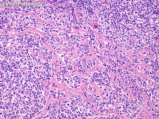
Microscopic Features: Primary Mediastinal Large B-cell Lymphoma (PMLBCL) (continued from previous image): Sclerosis is common and ranges from delicate collagen fibrils surrounding individual cells or groups of tumor cells to fibrous septa dividing tumor tissue into compartments. Fibrous septa are not as broad or thick as in classic Hodgkin lymphoma (cHL), nodular sclerosis type. Rare cases show features intermediate between PMLBCL and cHL and such cases have been separately grouped into so-called grey-zone lymphomas. Remnant of thymus with atrophy, hyperplasia, or cystic change may be seen. This image shows thin, delicate sclerotic bands surrounding small and large groups of tumor cells.
slide 5 of 21