
Pheochromocytoma : Immunohistochemical profile

slide 41 of 60


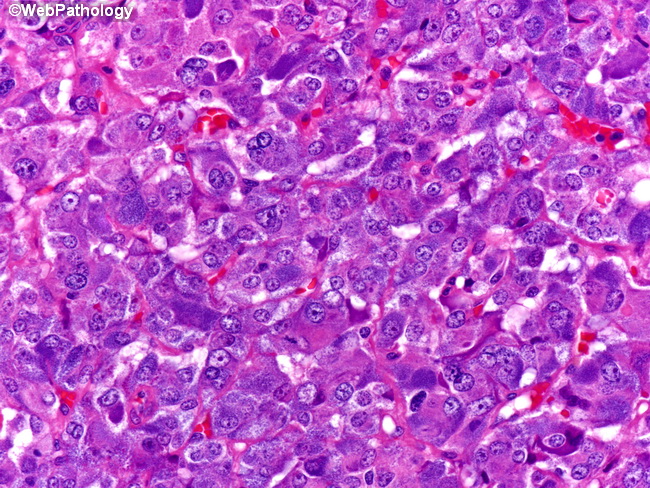
Comments:
Pheochromocytoma cells have finely granular basophilic or amphophilic cytoplasm. With electron microscopy, the cells show numerous dense-core neurosecretory granules containing catecholamines and other peptides. Immunohistochemically, they are positive for catecholamines, NSE, chromogranin, and synaptophysin. They may also be reactive for neurofilaments, serotonin, calcitonin, gastrin, vasoactive intestinal peptide, corticotropin and numerous other neuronal markers. Other markers that are positive in some cases include cytokeratin, vimentin, and HMB-45. The sustentacular cells surrounding tumor cell nests stain with S-100 protein immunostain (see image 35).
slide 41 of 60