
Pheochromocytoma

slide 37 of 60


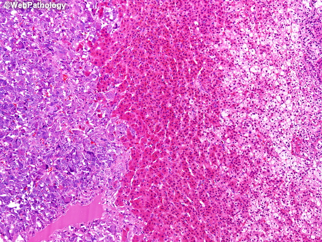
Comments:
Higher magnification of the previous image. Pheochromocytoma forms the left one-third of the image. The middle band of cells with deeply eosinophilic cytoplasm is zona reticularis. The right one third with vacuolated cytoplasm is zona fasciculata. Zona fasciculata and zona reticularis of the adrenal cortex synthesize glucocorticoids and sex hormones.
slide 37 of 60