
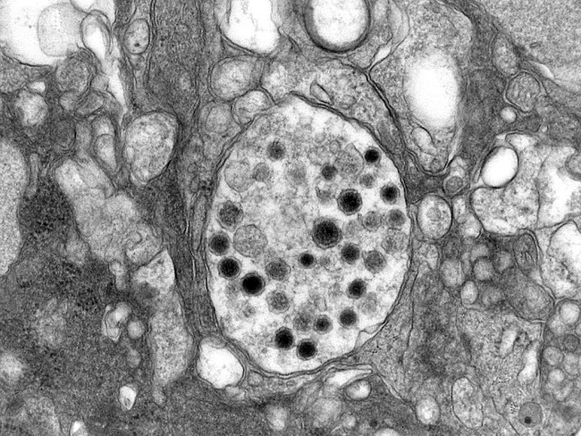
Neuroblastoma : Electron Microscopy

slide 34 of 60


Comments:
Higher magnification of the previous image showing a synaptic vesicle in a neuroblastoma cell. These vesicles are larger and more irregular than the normal presynaptic terminals. Numerous dense core neurosecretory granules are present. Courtesy of: Dr. Luciano de Souza Queiroz, Dept. of Pathology, Faculty of Medical Sciences, State University of Campinas (UNICAMP), Campinas, S�o Paulo State, BRAZIL. Additional images are here.
slide 34 of 60