
Branchial Cleft Cyst


Comments:
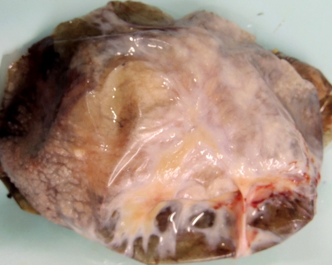
The photograph shows the inner wall of a branchial cleft cyst (same case as the previous two images). The inner surface appears irregular and granular (especially well seen in the lower left of this image) due to the presence of numerous hyperplastic lymphoid follicles. The branchial cleft anomalies include cysts, sinuses, fistulas as well as islands of cartilage. They are located in the anterolateral region of the neck and their exact location depends upon the specific branchial cleft involved. The first branchial pouch anomalies are rare and are located in the preauricular area or near the angle of the mandible. They may extend to the external auditory canal. Anomalies related to the second branchial pouch account for majority of the branchial cleft cysts. They are located mid-neck along the anterior border of sternocleidomastoid muscle. They may drain via a sinus tract into the pharynx near tonsillar fossa. Anomalies related to the third and fourth branchial clefts are rare. They are located in the lower neck in suprasternal or supraclavicular location. Case courtesy of: Dr. Sanjay D. Deshmukh, Professor of Pathology, Dr. Vithalrao Vikhe Patil Medical College & Hospitals, Ahmednagar, India.