
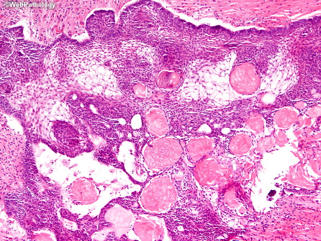
Adamantinomatous Craniopharyngioma

slide 15 of 32


Comments:
This image shows most of the key diagnostic features of an adamantinomatous craniopharyngioma: a) sheets of squamous epithelial cells with peripheral palisading (which is best seen along the top of the image); b) a loose meshwork of epithelial cells called the stellate reticulum; c) nodules of anucleated squames (ghost cells) with brighly eosinophilic cytoplasm termed wet keratin. Wet keratin is considered diagnostic even in the absence of viable epithelium.
slide 15 of 32